Diagnostic Tools
Visibly Better Eye Care uses state-of-the-art diagnostic tools at both our Lake Zurich and Mettawa locations. Read below to learn more about the instruments and technology we use to ensure the highest level of care for our patients.
- OPTOMAP ULTRA-WIDE FIELD RETINAL IMAGING SYSTEM
-
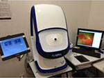
 Located in both our Lake Zurich and Mettawa locations is the Optomap Ultra-wide Field Retinal Imaging System. When you get an optomap image it’s fast, painless and comfortable. Nothing touches your eye at any time, making it suitable for the whole family. All you do is simply look into the device one eye at a time (like looking through a keyhole) and you will see a quick flash of light that tells you the image of your retina has been taken. The optomap ultra-widefield retinal image is a unique technology that captures more than 80% of your retina in one panoramic image while traditional imaging methods typically only show 15% of your retina at one time.
Benefits of an Optomap:
The benefits of having an optomap ultra-widefield retinal image taken include:
- Optomap facilitates early protection from vision impairment or blindness
- Early detection of life-threatening diseases like cancer, stroke, and cardiovascular diseaseThe unique optomap ultra-widefield view helps your optometrist detect early signs of retinal disease more effectively and efficiently than with traditional eye exams. Early detection leads to successful treatments that can be administered earlier and reduces the risk to your sight and health.
- ZEISS CIRRUS RETINAL SCANNER
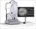
 Being able to analyze your eyes from multiple HD 3-D views provides comprehensive insight and analysis. And this benefits you in several ways:
- Spot small areas of pathology. Tightly spaced scans in the scanner ensure that small areas of pathology are imaged.
- Visualize the fovea. Scans that are spaced further apart than in the CIRRUS cube may miss the central fovea (a small depression in the retina of the eye where visual acuity is at its highest. The center of the field of vision is focused in this region, where retinal cones are particularly concentrated).
- Fuel for analysis. Millions of data points from the scanner are fed into the Zeiss proprietary algorithms for accurate segmentation, reproducible measurements and registration for change analysis over time.
- Take the pressure off the operator. As long as the scan is placed in the vicinity of the fovea or optic nerve, the retinal scanner automatically centers the measurements after the capture.
- See the tissue from different perspectives. The Optometrist is able to view the data from all angles, with 3D rendering, OCT fundus images and Advanced Visualization™.
- Future ready. Previously captured CIRRUS scans can be analyzed using new analyses.
The Zeiss CIRRUS Retinal Scanner is located in our Mettawa location.
- AUTO REFRACTOR
 Refraction testing is done as part of an eye examination for people who already wear glasses or contact lenses, but it can also be done if the results of the other visual acuity tests show that your eyesight is below normal and can be corrected by glasses. The Auto Refractor enables the doctor to make any necessary adjustments to ensure an accurate prescription for your glasses and/or contact lenses.
- AUTOMATED VISUAL FIELD ANALYZER*
 We have the Humphrey FDT perimeter that provides a clinically verified, fast and affordable means of detecting early visual field loss that can be caused by glaucoma, lesions inside the visual field pathway, and other diseases such as brain tumors.
The doctor will assess the focusing and eye tracking ability (this is especially important with children) Poor tracking and focusing can add to or be the cause of poor school performance.
*Additional fees apply.
- BIO-MICROSCOPE EXAMINATION (SLIT LAMP)
-
 A high powered magnifying lens enables the doctor to evaluate the structures around, inside, and in the back of the eye. Depending on the circumstances, doctor may wish to dilate your eye during your appointment, or at a later, more convenient time. This procedure more specifically checks for:
- Eye Infections
- Corneal Degeneration or Injury
- Blepharitis (dandruff on the eyelashes
- Blocked Tear Ducts
- Styes
- Dry Eye Evaluation (tear assessment)
- Cataracts
- Glaucoma
- Pupil Anomalies
- Macular Degeneration
- Optic Nerve Health
- General Retinal Health
- TONOMETRY
 This device is used for the ‘Puff” test which measures the fluid pressure inside the eye. It is an important test in the detection and evaluation of glaucoma.
Call Visibly Better Eye Care today for a complete vision evaluation!

